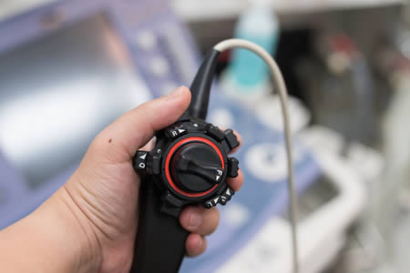
Transesophageal echocardiogram (TEE) is an echocardiogram (i.e. a test which uses an ultrasound beam to obtain images of the heart) for which images are obtained via the esophagus. As the esophagus passes directly behind the heart, this type of echocardiography can be used for specific indications to obtain better images than standard (transthoracic) echocardiography.
 The TEE probe is a long flexible tube which is placed into the back of the throat and is then swallowed. Once in the esophagus, images are obtained in real time and interpreted by your doctor. It may take up to 10-15 minutes for image acquisition.
Common utilization of TEE include: evaluation for presence of clot (or thrombus) prior to certain procedures (e.g. cardioversion or ablation); identify potential causes of stroke; detailed evaluation of valve issues or for the presence of infection on a heart valve; or to evaluate for the presence of a “hole” between the top chambers of the heart (patent foramen ovale or atrial septal defect). A TEE may also be considered if transthoracic echo is inadequate.
In general, TEE may be done as an outpatient procedure under conscious sedation via an IV. As you will be receiving sedation, you will need to arrange for a ride home. You will be asked not to eat or drink for at least 8 hours prior to your scheduled procedure time. Further more detailed instructions will be provided upon scheduling.

